Classify oral mucus membrane. Write in detail about histology of gingiva.

DENTAL ANATOMY, EMBRYOLOGY AND ORAL HISTOLOGY

🔍 LONG ANSWERS:

Q: Classify oral mucus membrane. Write in detail about histology of gingiva. (15marks)

Ans.

💡Classification of Oral Mucous Membrane

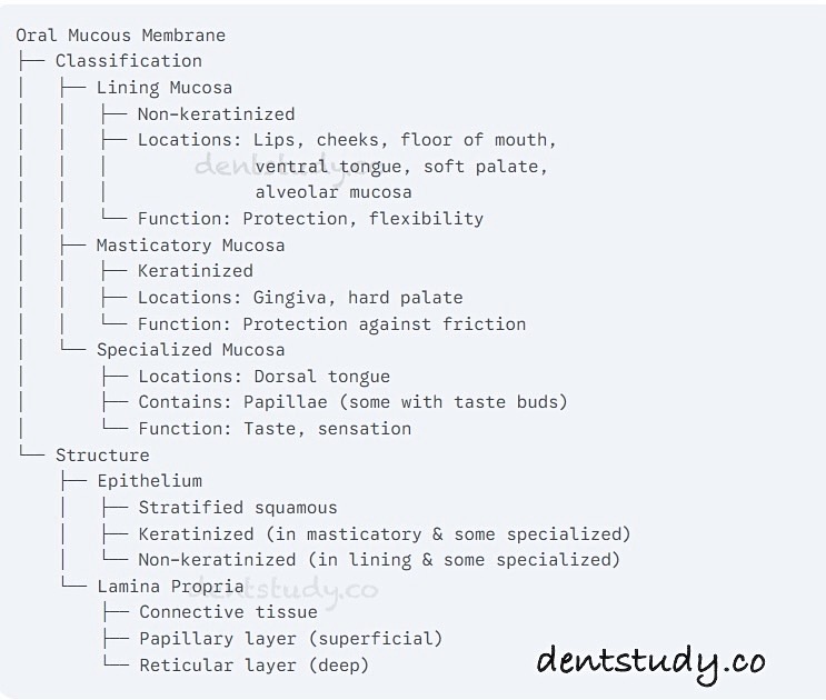
The oral mucosa lines the inside of the oral cavity and is classified into three main types based on function and structure:

1. Masticatory Mucosa: Found in areas subject to friction and pressure during chewing, such as the gingiva and hard palate. It is keratinized, providing a tough, protective surface.

    • Location: Gingiva and hard palate.

    • Characteristic: Keratinized or parakeratinized epithelium.

    • Function: Withstands mechanical stress during chewing.

2. Lining Mucosa: This type covers the inner surfaces of the lips, cheeks, floor of the mouth, underside of the tongue, soft palate, and alveolar mucosa. It is non-keratinized, providing flexibility and allowing for movement.

    • Location: Lips, cheeks, floor of the mouth, ventral surface of the tongue, soft palate, alveolar mucosa.

    • Function: Allows flexibility and movement.

    • Characteristic: Non-keratinized epithelium.

3. Specialized Mucosa: Located on the dorsum of the tongue, it contains specialized structures called papillae, some of which house taste buds.

    • Location: Dorsal surface of the tongue.

    • Function: Contains taste buds for taste perception.

    • Characteristic: Keratinized or non-keratinized epithelium with specialized papillae.

💡Histology of Gingiva


🔸 The gingiva is the part of the masticatory mucosa that surrounds the necks of the teeth and covers the alveolar bone. Histologically, gingiva is part of the masticatory mucosa and consists of epithelial and connective tissue layers.

  • Epithelium: Stratified squamous keratinized epithelium, which can be further divided into:
    • Oral epithelium: Faces the oral cavity and is keratinized or parakeratinized.
    • Sulcular epithelium: Lines the gingival sulcus (the space between the tooth and gingiva) and is non-keratinized.
    • Junctional epithelium: Attaches the gingiva to the tooth enamel, forming a seal. It is non-keratinized.
  • Lamina propria: Connective tissue underlying the epithelium, composed of:
    • Papillary layer: Connects to the epithelium with connective tissue papillae.
    • Reticular layer: Deeper layer that blends with the periosteum of the alveolar bone.

🔸The gingiva is further divided into:

  • Marginal gingiva (free gingiva): Unattached portion forming the gingival sulcus.
  • Attached gingiva: Firmly bound to the underlying alveolar bone and cementum.
  • Interdental gingiva (gingival papilla): Fills the space between adjacent teeth.

1. Epithelial Layer

  • The gingival epithelium is stratified squamous epithelium, which can be:

    • Keratinized: In the free and attached gingiva.

    • Non-keratinized: In the sulcular epithelium.

  • Junctional epithelium: A specialized non-keratinized epithelium that attaches the gingiva to the tooth surface via hemidesmosomes.

Types of Gingival Epithelium

  • Oral Epithelium: Faces the oral cavity, keratinized.

  • Sulcular Epithelium: Lines the gingival sulcus, non-keratinized.

  • Junctional Epithelium: Forms the base of the gingival sulcus, highly permeable.

2. Connective Tissue (Lamina Propria)

  • Composed of fibroblasts, collagen fibers (Type I), blood vessels, and nerves.

  • Gingival Fibers:

    • Dentogingival fibers: Connect the gingiva to the tooth.

    • Circular fibers: Encircle the tooth.

    • Transseptal fibers: Connect adjacent teeth.

3. Blood Supply

  • Supplied by branches of the superior and inferior alveolar arteries.

4. Nerve Supply

  • Innervated by branches of the trigeminal nerve (cranial nerve V).

5. Cell Types in Gingival Epithelium

  • Keratinocytes: Predominant cells involved in keratin production.

  • Melanocytes: Pigment-producing cells.

  • Langerhans Cells: Part of the immune defense.

  • Merkel Cells: Sensory cells.

👉 Summary:

  • Gingiva, being a part of masticatory mucosa, is primarily keratinized, except in sulcular and junctional areas.

  • The epithelial layer protects against mechanical forces, while the connective tissue provides support and nourishment.

  • Blood vessels, nerves, and fibers play crucial roles in maintaining gingival health.

BRAIN MAP FOR ORAL MUCOUS MEMBRANE: