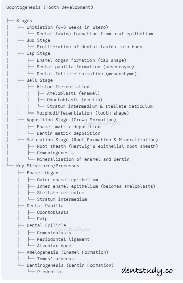
Essay about tooth development. Write note on histological and morphological differentiation of odontogenesis.

DENTAL ANATOMY, EMBRYOLOGY AND ORAL HISTOLOGY

πŸ”Ž Long EssayΒ 

Q. Write an essay about tooth development. Add a note on histological and morphological differentiation of odontogenesis.

Ans.

πŸ“Œ Tooth Development (Odontogenesis)

πŸ‘‰ Tooth development, also known as odontogenesis, is a complex, highly regulated process that begins early in embryonic life and continues through early childhood. It involves interactions between the oral epithelium and the underlying ectomesenchyme, leading to the formation of primary and permanent teeth. The process can be divided into several stages, each marked by distinct morphological and histological changes.

πŸ‘‰ Tooth development, or odontogenesis, is a complex process involving reciprocal interactions between oral epithelium and underlying mesenchyme, leading to the formation of teeth. This process is conventionally divided into several overlapping stages: initiation, bud, cap, bell, apposition, and maturation.

πŸ‘‰ Tooth development, or odontogenesis, is a complex process involving interactions between oral epithelium and underlying mesenchymal tissue. It occurs in several stages:

πŸ”ΈInitiation stage: Oral epithelium proliferates to form the dental lamina.

πŸ”ΈBud stage: Dental lamina forms bud-like structures that will develop into teeth.

πŸ”ΈCap stage: Bud invaginates, forming a cap shape with enamel organ, dental papilla, and dental follicle.

πŸ”ΈBell stage: Enamel organ differentiates into ameloblasts (enamel-forming cells), and dental papilla differentiates into odontoblasts (dentin-forming cells).

πŸ”ΈApposition stage: Enamel and dentin are laid down in layers.

πŸ”ΈMaturation stage: Enamel and dentin undergo mineralization.

➑️Stages of Tooth Development

  1. Initiation (Bud Stage)

    • Timeline: Begins at 6–7 weeks of intrauterine life.
    • Morphological Features: Formation of a localized epithelial proliferation called the dental lamina into the underlying mesenchyme.
    • Histological Features: Rounded clusters of proliferating epithelial cells surrounded by ectomesenchymal cells.
  2. Proliferation (Cap Stage)

    • Timeline: 8th week of gestation.
    • Morphological Features: The enamel organ forms a cap-like structure with an underlying dental papilla and surrounding dental sac.
    • Histological Features: The enamel organ contains:
      • Outer enamel epithelium: Protective layer.
      • Inner enamel epithelium: Precursor to ameloblasts.
      • Stellate reticulum: Star-shaped cells providing structural support.
  3. Differentiation and Morphogenesis (Bell Stage)

    • Timeline: 9–10 weeks of gestation.
    • Morphological Features: The enamel organ assumes a bell shape. The dental papilla forms the pulp, and the dental sac develops into supporting structures (periodontium).
    • Histological Features:
      • Stratum intermedium appears between the inner enamel epithelium and stellate reticulum, aiding in enamel production.
      • Ameloblasts and odontoblasts differentiate, initiating hard tissue formation.
  4. Apposition Stage

    • Timeline: Begins shortly after the bell stage.
    • Morphological Features: Deposition of dentin and enamel.
    • Histological Features:
      • Odontoblasts secrete predentin, which mineralizes to form dentin.
      • Ameloblasts deposit enamel over the dentin.
  5. Maturation Stage

    • Enamel and dentin undergo full mineralization.
    • Root formation and eruption follow.

πŸ“Œ Histological and Morphological Differentiation of Odontogenesis

πŸ”ΉHistological differentiation: Involves the differentiation of cells into specialized tissues:

    • Amelogenesis: Formation of enamel by ameloblasts.
    • Dentinogenesis: Formation of dentin by odontoblasts.
    • Cementogenesis: Formation of cementum by cementoblasts.
    • Periodontal ligament formation: Formation of periodontal ligament by fibroblasts.

πŸ”ΉMorphological differentiation: Involves the shaping of the tooth:

    • Crown formation: Determined by the shape of the enamel organ.
    • Root formation: Occurs after crown formation, involving Hertwig’s epithelial root sheath.

Β 

✳️Histological Differentiation in Odontogenesis

Histological differentiation refers to the transformation of cells into specialized forms during tooth development:

  1. Ameloblast Differentiation:

    • Cells of the inner enamel epithelium elongate into columnar ameloblasts responsible for enamel formation.
    • Ameloblasts pass through presecretory, secretory, and maturation phases to produce enamel.
  2. Odontoblast Differentiation:

    • Cells of the dental papilla differentiate into odontoblasts, secreting dentin matrix.
    • Odontoblasts retreat pulpward, leaving dentinal tubules.
  3. Cementoblast Differentiation:

    • Dental sac cells differentiate into cementoblasts that form cementum.

✳️ Morphological Differentiation in Odontogenesis

Morphological differentiation involves the shaping of the tooth:

  • Crown Shape Formation: The shape of the crown (incisor, molar, etc.) is determined during the bell stage by folding of the inner enamel epithelium.
  • Root Formation: Begins after crown development; guided by Hertwig’s epithelial root sheath, which determines the root’s length and shape.

ODONTOGENESIS (TOOTH DEVELOPMENT)